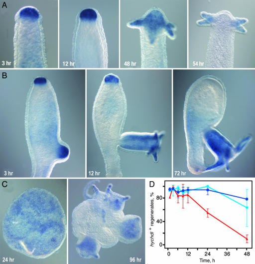
Fig. 3.
Expression of hychdl during regeneration and reaggregation. hychdl expression during regeneration in wild-type animals (A) and in the regeneration-deficient mutant strain reg-16 (B). Note the strong up-regulation of hychdl expression in apical tissue despite failure of head regeneration. (C) Expression of hychdl in reaggregates. Initially uniform endodermal staining becomes restricted to newly formed heads. (D) Kinetics of up-regulation of hychdl during regeneration. Percentage of hychdl-positive regenerates at different time points after bisection was analyzed in three independent experiments with >11 regenerates. No significant difference can be seen during head regeneration after bisection at 50% BL (light blue) and 80% BL (dark blue); red, foot regeneration (50% BL).

