Fig. 2.
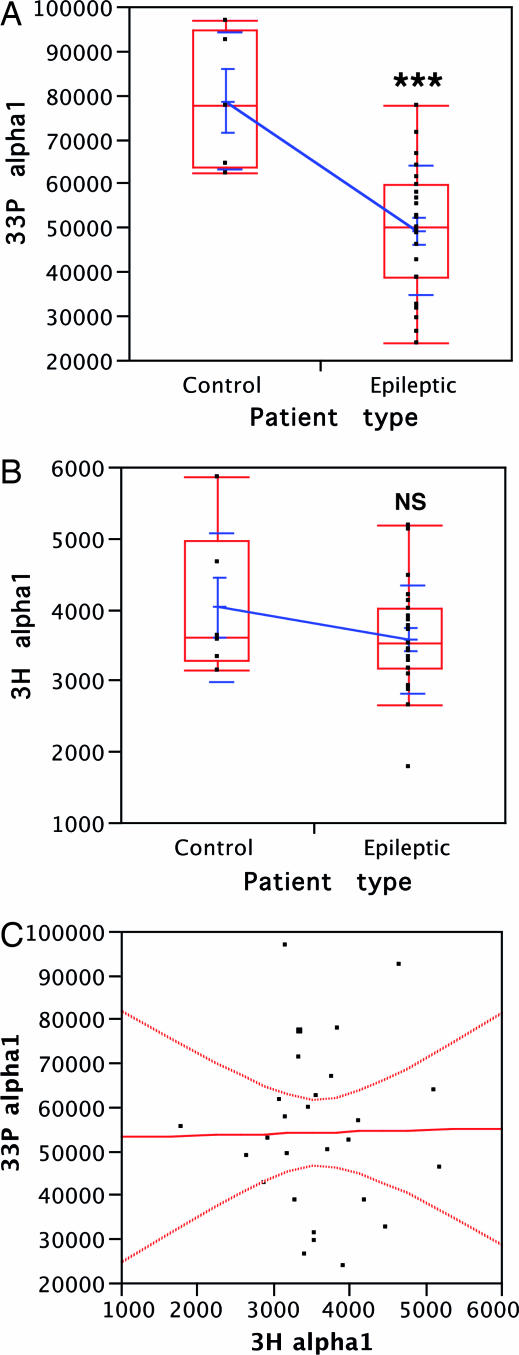
The deficiency of GABAAR endogenous phosphorylation in membranes from epileptogenic tissue is intrinsic. (A) 33P-labeled phosphate incorporation (in counts) into α1-subunit by the endogenous phosphorylation compared between nonepileptogenic cortex (n = 5) and epileptogenic cortex (n = 23). The distributions are presented as mean values with SEM and SD (blue) and as median values in quartile boxes (red). ∗∗∗, P < 0.001, Student's t test. (B) 3H-incorporation (in dpm) in α1 by the [3H]flunitrazepam labeling compared between nonepileptogenic cortex (n = 6) and epileptogenic cortex (n = 23). Distributions are presented as in A. NS, nonsignificant (P = 0.24 for the t test). (C) Correlation graph between the individual 33P- and 3H-labeling values of membranes from surgical samples of both epileptic and nonepileptic patients (n = 27). The calculated correlation r = 0.014 and the null leverage indicated the absence of interaction between the two labelings (P = 0.95).

