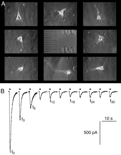
Fig. 3.
Rundown of GABAA currents measured by whole-cell patch clamp on acutely dissociated neurons from cortical tissue of surgery patients. (A) Phase-contrast micrograph of isolated pyramidal neurons. The scale is in millimeters with small graduations of 10 μm. (B) Current recording traces of a neuron from an epileptic patient. GABA (100 μM) was applied for 1 s every 3 min.

