
Fig. 5.
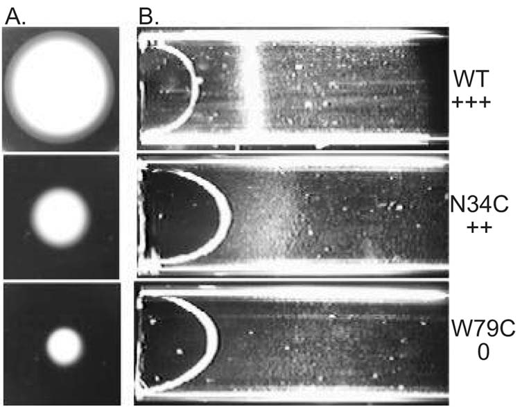
Comparison of aerotaxis responses in succinate semi-soft agar (A), and a capillary (B). A. Cells (5 μl) in mid-logarithmic phase were inoculated in the center of a succinate semi-soft agar plate (H1 medium, 30 mM succinate, 0.28% agar, 100 μg/ml ampicillin) and incubated at 30º C for 24 hours. Cells with a wild type aerotaxis phenotype formed a distinct convex ring (top) that is absent in mutants that have a null phenotype for aerotaxis (bottom). Other mutants were partially defective in aerotaxis (center). B. Cells (O.D.600 = 0.45) in Luria-Bertani medium were loaded into an optically flat capillary. Band formation is shown for each of the strains used in (A).