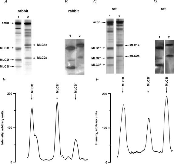
Figure 1. SDS-PAGE (A and C), Western blots (B and D) and the densitometry (E and F) of myosin light chain isoforms of rabbit and rat skeletal muscle.
The figure shows silver-stained 10–20% gradient SDS-PAGE (A and C) and Western blots (B and D) of single fibres. Lanes 1 in A and C are electrophoretograms of type IIB fibres containing exclusively fast MLC isoforms (MLC1f, MLC2f, MLC3f). Likewise, lanes 2 in A and C are electrophoretograms of type I fibres containing exclusively slow MLC isoforms (MLC1s, MLC2s). Based on the reactivity with a monoclonal antibody against alkali MLCs and a monoclonal antibody against regulatory MLCs (Western blots B and D) the myosin light chain isoforms were identified as labelled in A and C. The traces in E and F are the densitometric profiles of lanes 1 in C (SDS-PAGE) and D (Western blot), respectively.

