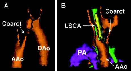
Figure 2.

Three-dimensional reconstruction of the arch anatomy of patient 7, the most common arch we encountered. Panel A is right posterior oblique view and panel B is left posterior oblique. Both views show the coarctation (Coarct) and ascending aorta (AAo), with the descending aorta (DAo) seen well in panel A. In panel B, the left subclavian artery (LSCA) is shown arising from a retroesophageal (Kommerell’s) diverticulum. In this same panel, the trachea is green, the esophagus is yellow, and the pulmonary artery (PA) is purple.
