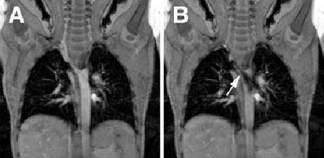
Figure 3.

Coronal cine-MRI of patient 7. Panel A is from a frame without significant flow across the area of coarctation. Panel B is later in the cardiac cycle demonstrating significant turbulence in the posterior descending aorta, arising from the area of narrowing (arrow).
