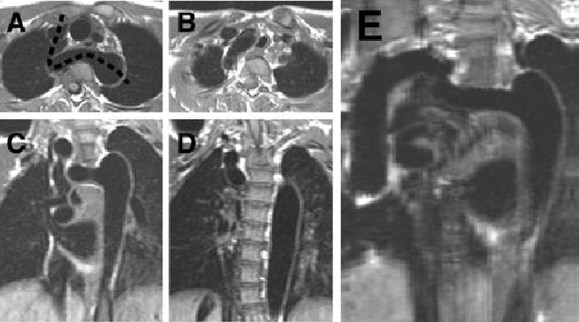
Figure 4.

Several views of the arch of patient 8, including a curved-cut MRI. Panels A and B are selected T1-weighted axial images, moving caudally to cranially. Panels C and D are T1-weighted coronal images, moving anteriorly to posteriorly. Panel E represents the reconstruction along the plane of the dashed line in panel A. The tortuous nature of the arch, as well as the elongated area of narrowing, is well seen in this reconstruction.
