
Figure 3. The autophagosome marker GFP-LC3 colocalizes with markers of MHC class II loading compartments in human epithelial cell lines and dendritic cells.
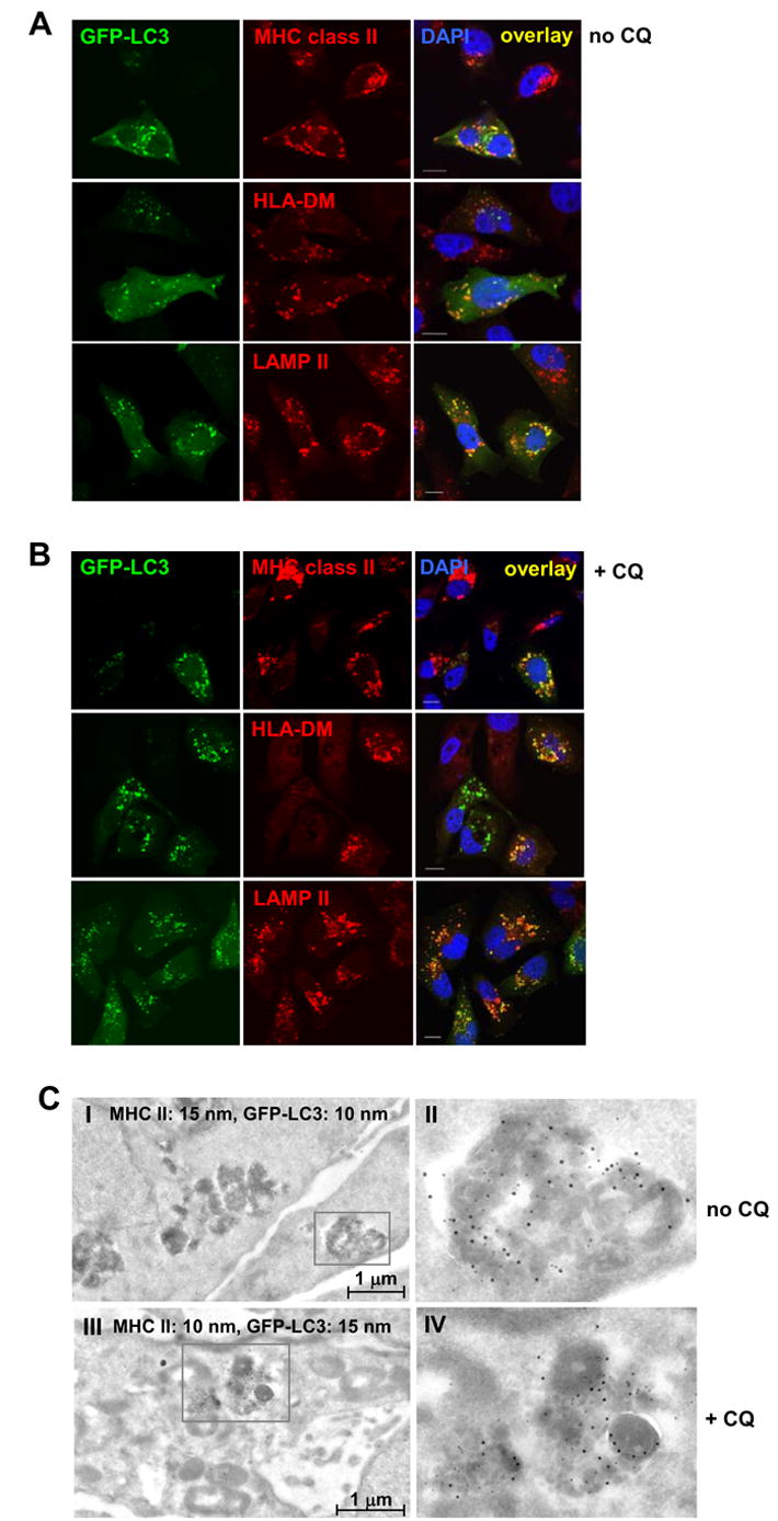
(A) MDAMC cells were treated with 200 U/ml IFN-γ, transiently transfected with a GFP-LC3 reporter construct and 48 h later stained with antibodies to MHC class II, HLA-DM, LAMP-2 and DAPI for DNA content. Colocalization of GFP-LC3 with MIIC markers was analyzed by confocal microscopy. Scale bars: 10 μm. Representative cells from one experiment out of three are shown.
(B) Same as (A), except that 50 μM chloroquine (CQ) was present during the last 10 h of the culture. Scale bars: 10 μm. Representative cells from one experiment out of three are shown.
(C) Colocalization of GFP-LC3 and MHC class II molecules in electron-dense multivesicular compartments. Untreated (I and II) or CQ-treated (III and IV) MDAMC cells stably expressing GFP-LC3 and MHC class II-positive due to IFN-γ induction were fixed in 4% paraformaldehyde and cut into 80 nm-thin cryosections. Sections were labeled with an HLA-DR-specific antiserum and 10 nm protein A-Gold (PAG10) and antibody-PAG complexes were irreversibly fixed with glutaraldehyde. Subsequently, sections were labeled with a GFP-specific antibody and 15 nm protein A-Gold (PAG15) and were analyzed by electron microscopy. As a control, PAG10 and PAG15 were interchanged and were shown to produce the same labeling pattern (I/II vs. III/IV). Insets from panels I and III are shown at higher magnification in panels II and IV, respectively. Representative fields from one experiment out of three are shown.