
Figure 3.
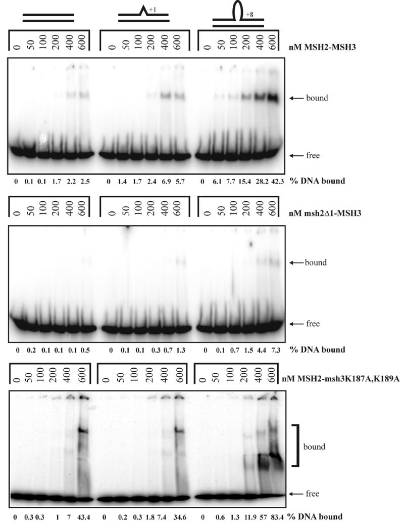
Gel mobility shift assays of MSH2-MSH3 (top panel), msh2Δ1-MSH3 (second panel), and MSH2-msh3K187A,K189A (bottom panel) incubated with homoduplex, +1 loop, and +8 loop substrates as described in the Materials and Methods. The percentage of labeled DNA substrate that was bound is indicated below each gel.