
Figure 10.
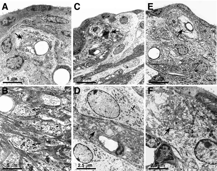
UItrastructural changes in type II fibrocytes after noise exposure. A. Zero hours after 100 dB exposure in the lower apical turn, degeneration of type II fibrocyte is seen in the spiral prominence (arrow). B. Twenty-four hours after exposure to 116 dB in the lower apical turn, cellular debris remains after lysis of type II fibrocytes (arrows). C. Eight hours after exposure to 116 dB in the upper basal turn, some type II fibrocytes are pyknotic with nuclear condensation (arrow). D. Twenty-four hours after exposure to 112 dB in the lower apical turn, mitochondrial swelling is seen within type II fibrocytes (black arrow); however, intervening root cells appear normal (white arrow). E. Two weeks after exposure to 116 dB in the lower basal turn, the spiral prominence shows loss of type II fibrocytes around the central vessel (arrow). F. Eight weeks after exposure to 116 dB in the lower apical turn, processes of type II fibrocytes are present in the spiral prominence (arrow), suggesting regeneration or repair of this cell type.