
Figure 5.
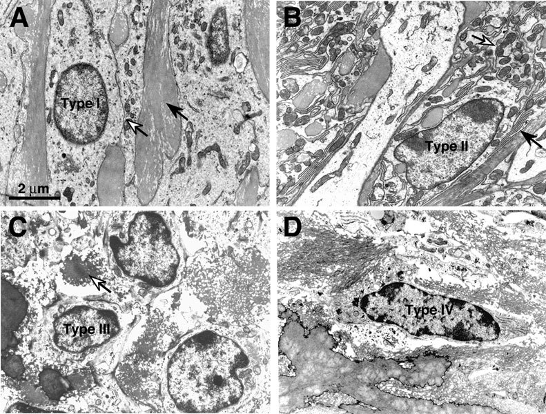
Ultrastructure of normal spiral ligament fibrocytes. A. Type I fibrocytes are electron-lucent, associated with collagen bundles (black arrow), and connected by gap junctions (white arrow). B. Type II fibrocytes are associated with root cells, rich in mitochondria (white arrow), and have extensive interdigitating processes (black arrow) with adjacent type II cells. C. Type III fibrocytes appear round in cross-section due to their circumferential orientation with respect to the cochlear spiral and are associated with spiraling collagen bundles (white arrow). D. Type IV fibrocytes are fusiform, contain scant granular cytoplasm, and do not have the extensive processes of the type II cells. Scale bar applies to A–D.