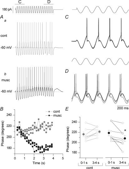
Figure 4. Muscarinic ADPs are associated with AP phase shift in O-LM interneurones.
A, representative voltage output in response to a 4 s, 90 pA sinusoidal current in the absence (a) or presence (b) of 10 μm muscarine. B, AP phase over the course of the 4 s train for 3 trials in both control (grey) and 10 μm muscarine (black). Expanded, overlaid traces during the first (C; 0–1 s in A) or last (D; 3–4 s in A) second of the sinusoidal current injection. While the APs occur at a similar time in the cycle initially, mAChR activation shifts the timing of AP generation to earlier in the cycle. E, phase for control (grey; n = 7) and upon mAChR activation (black; n = 8) in the first (0–1) or last (3–4) second. In control conditions, the phase increased from 212 ± 3 to 220 ± 3 degrees (6 of 7 cells, P = 0.03; P = 0.13 for all 7 cells). Concomitant with the development of a muscarinic ADP (by 1.7 ± 0.5 mV, P = 0.01, n = 10), mAChR activation shifted phase from 218 ± 6 to 208 ± 6 degrees (n = 8, P = 0.026). The ADP was measured by comparing the difference in average voltage in seconds 0–1 versus 3–4 of the sinusoidal stimulation; the sinusoidal stimulation itself generated no net voltage difference in control conditions (see Methods).

