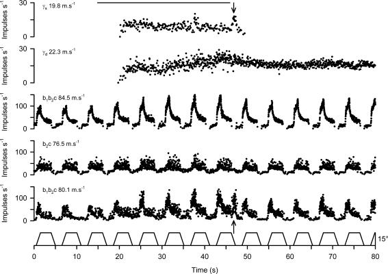
Figure 1. Identification of static and dynamic γ axons in TA.
The records from above downward are the stimulus signal, instantaneous frequency of two γ axons, and three muscle spindle afferents and the TA stretch waveform (ankle angle). The stimulus was a train of pulses (0.2 ms, 10 Hz, 2.0 V) delivered dorsal to the cuneiform nucleus on the right hand side. The conduction velocities of all the axons are shown inset on the left. The spindle primary afferents are designated as b1b2c or b2c as found by succinylcholine testing. The arrows indicate the burst of firing in the lowermost spindle coinciding with a burst in the upper γ axon.

