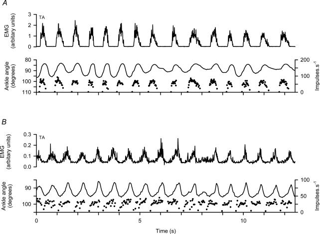
Figure 6. Patterns of firing of γd axons during spontaneous locomotion.
A and B, data from two different experiments. Each shows, from above downwards, TA EMG, ankle rotation and instantaneous frequency of a TA γd axon. Note in each cycle the rapid onset of firing from a silent background and the relative constancy during muscle shortening.

