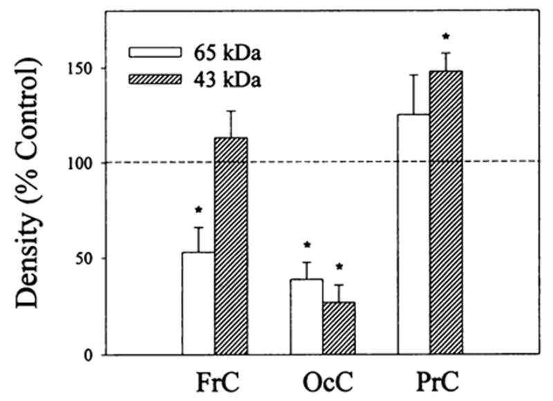
Fig. 5.

Densitometric analysis of 125I-β-endorphin cross-linked bands in frontal, parietal and occipital cortex membrane preparations from rats treated with buprenorphine. Rats were administered (i.p.) 2.5 mg/kg buprenorphine; 20 hr later they were sacrificed and their brains were removed and dissected. Membrane preparations (after five washes with 50 mM Tris-HCI, pH 7.4) were cross-linked with 2 nM 125I-β-endorphin in the presence (nonspecific binding) and absence (total binding) of 1 μM unlabeled β-endorphin as described in “Materials and Methods.” Equal amounts of protein (150–200 μg) were solubilized in sodium dodecyl sulfate (SDS) buffer and polyacrylamide gel ectrophoresis (PAGE) (10%) was performed. Specific densities of the 60- to 65- and 43- to 46-kDa bands in each brain region were determined as the difference between the density of the total and nonspecific binding for control and buprenorphine treated samples. Observed density changes induced by the buprenorphine treatment are expressed as percent of control values that were normalized to 100%. Data are the mean ± S.E.M. of three to four experiments, *vs. controls, P < .05.
