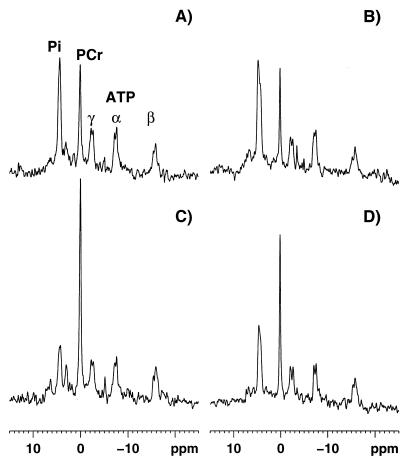
Figure 1.
Calf muscle 31P-MRS spectra. (Upper) Spectra (eight free induction decays) from a normal subject (A) and patient 2 (B) at the end of in-magnet work. (Lower) Spectra (eight free induction decays) collected between 16 and 32 sec of recovery from the same subjects (C, normal subject; D, patient 2). During incremental exercise there is a progressive reduction of the PCr peak, hydrolyzed via the creatine kinase reaction to buffer ATP concentration, and an increase in the Pi peak produced by the ATP hydrolysis during muscle contraction (A and B). As soon the exercise is stopped PCr and Pi concentrations begin to return to their pre-exercise values: PCr is resynthesized from ATP, via the creatine-kinase reaction, and Pi is used for ATP synthesis (C and D). The ATP production during recovery from exercise is entirely caused by oxidative phosphorylation (10), thus the PCr resynthesis rate reflects precisely the mitochondrial rate of ATP production. At the same recovery time point, the PCr peak is smaller in patient 2 (D) than in the control subject (C), indicating a reduced rate of muscle mitochondrial ATP production in the FRDA patient. The abscissa reports the chemical shift in ppm and ordinate the relative signal intensity in arbitrary units.

