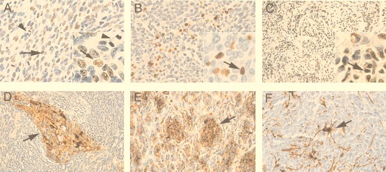
Figure 4.
Immunohistological analysis of human of medulloblastomas. Immunostaining of tumor samples with anti-T antigen antibody (A, B, and C). Arrows indicate T antigen-positive cells. Arrowhead in A and A Inset show the presence of mitotic figures. Immunostaining of tumor samples with neuronal markers including synaptophysin (D), class III β-tubulin (E), and glial fibrillary acidic protein (GFAP, F) is shown [hematoxylin counterstain, original magnification: ×400 (A, B, E, and F); ×200 (C and D); ×1,000 (Insets)].

