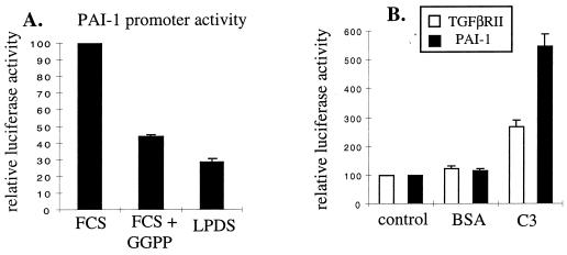
Figure 6.
Regulation of TGFβ signaling via the geranylgeranylation pathway. (A) Cells were transfected with p3TP-Lux plus pCMVβgal. After recovery, cells were incubated for 16 h in media with: lane 1, FCS alone; lane 2, FCS plus 10 μM GGPP; lane 3, LPDS alone. Luciferase activity was normalized to β-galactosidase activity. Data are plotted as the mean ± SEM of three independent experiments. (B) Cells were transfected with either pTGFβRII-500/36-Lux or p3TP-Lux plus pCMVβgal. After recovery, cells were cultured for 16 h in media supplemented with LPDS plus: lane 1, control; lane 2, 50 μg BSA/ml; lane 3, 50 μg C3 exotoxin/ml. Luciferase activity was normalized to β-galactosidase activity. Values are the mean ± SEM of three independent experiments.

