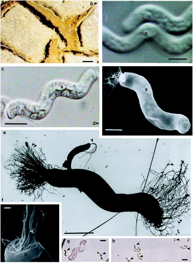
Figure 1.
(a) Mat surface at the Ebro Delta field site (3) showing lack of standing water. (Bar = 10 cm.) (b) Two spirilla cells (S, sulfur globule) shown by differential interference contrast (Nomarski). (Bar = 5 μm.) (c) Phase contrast microscopy of live spirillum cells. (Bar = 5 μm.) (d) Bipolar lophotrichous large spirillum in which only one pole has retained flagella. Sulfur globules are visible through the cell wall (scanning electron micrograph). (Bar = 5 μm.) (e) Negative-stain transmission electron micrograph of an entire bipolar lophotrichous large spirillum showing flagella “braids” (double arrowheads) compared with standard-sized spirilla (single arrowhead). (Bar = 5 μm.) (f) This scanning-electron micrograph of a cell terminus shows one vaulted end with residual flagella. The indentation coated by the polar organelle (P; see Fig. 2) is implied. (Bar = 0.5 μm.) (g) This Gram-stain brightfield preparation compares the two size classes, huge and standard, of Gram-negative spirilla. (Bar = 5 μm.) (h) Standard-sized spirillum Gram stain. The lighter spots are probably sulfur globules. (Bar = 5 μm.)

