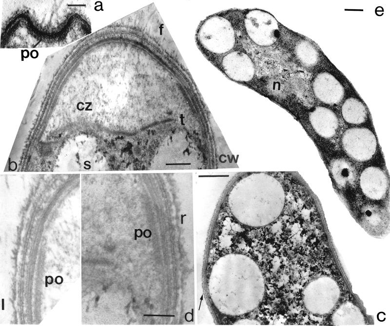
Figure 2.
Transmission electron micrographs of Titanospirillum velox. (a) The polar organelle (po) underlies the indented terminus. (Bar = 1 μm.) (b) Complex cell wall (cw), flagella (f), and clear zone (cz) at the cell terminus subtended by thickening (t) and empty sulfur-globule (s) vacuoles. (Bar = 0.5 μm.) (c) Distended cell wall around a peripheral membrane-bound sulfur-globule vacuole (arrow). (Bar = 1 μm.) (d) The left (l) and right (r) polar organelles lie proximal to at least nine layers of wall material at the cell termini. (Bar = 0.25 μm.) (e) Sulfur-globule vacuoles distributed irregularly in the cytoplasm are especially abundant at the cell periphery distal to the nucleoid (n). (Bar = 1 μm.)

