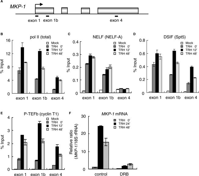
Figure 2.
Association of pol II, NELF, DSIF and P-TEFb with the MKP-1 gene. (A) Rat MKP-1 genomic locus with the primer positions. (B–E) Distributions of pol II and elongation factors on the MKP-1 gene. ChIP assay was performed with an anti-pol II (N-20) (B), an anti-NELF-A (C), an anti-Spt5 (D) and an anti-cyclin T1 antibody (E) with chromatin prepared at various time points of TRH stimulation. Density on the MKP-1 gene was quantified by real-time PCR and presented as a percentage of input. A typical experiment (mean ± SD, n = 3) repeated three times is shown. (F) Suppression of MKP-1 transcription by DRB an inhibitor of P-TEFb. Cells were incubated with or without 30 μM DRB for 2 h prior to TRH stimulation. Total RNA was extracted 24 and 48 min after TRH, and transcripts of the MKP-1 were quantified by real-time PCR. A typical experiment repeated three times (mean ± SD in triplicates) is shown.

