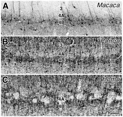
Figure 4.
NPNF immunostaining in layer 4A as seen with differential-interference contrast optics. In Macaca (A) and Pongo (B), layer 4A is generally lightly stained, although some vertically oriented dendrites and cell bodies are present that stain darkly for NPNF. By contrast, in Homo (C), layer 4A is laced with bands of dark, NPNF-immunoreactive tissue that encapsulate pockets of lightly stained tissue. (Bar = 100 μm.)

