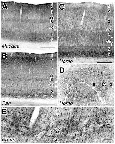
Figure 5.
MAP 2 immunostaining of area V1 in Macaca (A), Pan (B), and Homo (C–E). A–C are from standard coronal and horizontal sections. D is a from a section that passes tangentially through the middle cortical layers in a human. E shows the organization of human layer 4A in the coronal plane with DIC optics. A distinctive, patchy, mesh-like pattern is apparent in layer 4A of humans in both coronal and tangential planes. [Bar = 500 μm (A–D) and 100 μm (E)].

