
Figure 3.
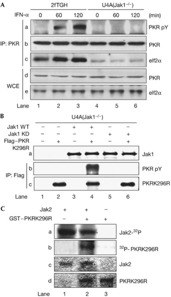
Tyrosine phosphorylation of PKR by Jaks. (A) 2fTGH and U4A cells were stimulated with IFN-α2b for the indicated time periods. Protein extracts were immunoprecipitated (IP) for PKR followed by immunoblotting with phosphotyrosine 4G10 antibody (panel a), PKR monoclonal antibody (panel b) and eIF2α antibody (panel c). The levels of PKR (panel d) and eIF2α (panel e) in WCE were detected by immunoblotting. (B) U4A cells were transfected with the catalytically inactive Flag–PKRK296R complementary DNA in the absence or presence of either wild-type (WT) or a kinase-dead (KD) K296R mutant of Jak1 cDNA. Protein extracts were immunoprecipitated for Flag followed by immunoblotting with phosphotyrosine 4G10 (panel b) or PKR antibody (panel c). Jak1 levels (panel a) were detected by immunoblotting of WCE. (C) Recombinant murine Jak2 and GST–PKRK296R were subjected to in vitro phosphorylation with [γ-32P]ATP and autoradiography (panels a and b). The phosphorylated proteins were detected after an equal time of exposure. Total Jak2 (panel c) and GST–PKRK296R (panel d) levels were detected by Coomassie blue staining. IFN, interferon; PKR, dsRNA-dependent protein kinase; PKRK296R, PKRLys296Arg; WCE, whole-cell extracts.