
Figure 2.
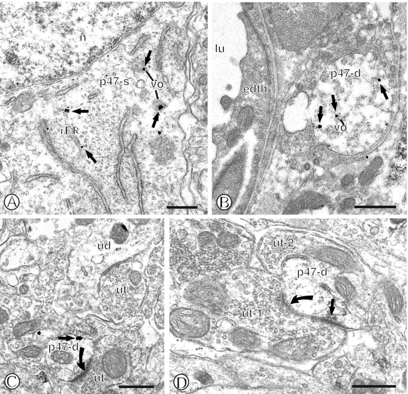
Immunogold labeling of p47phox in mNTS neurons. (A). Immunogold particles (small closed arrows) for p47phox are present in diverse intracellular compartments of a single labeled soma (p47-s). Gold-silver deposits can be seen near rough endoplasmic reticula (rER) just beneath the nucleus (n), as well as nearby small vesicular organelles (vo). (B). A single immunogold labeled dendritic profile (p47-d) contains gold labeling for p47phox (small closed arrow) near vesicular organelles (vo) and the cytoplasm. This dendritic profile is present in proximity to the neurovasculature as indicated by a vessel lumen (lu) and an associated endothelial cell (edth). (C). A single gold labeled small dendritic profile (p47-d) is present within the neuropil. This dendritic profile contains immunogold labeling on the extrasynaptic surface membrane adjacent to an unlabeled axon terminal (ut) that forms an excitatory-type asymmetric synapse (closed curved arrow). Unlabeled dendritic (ud) and axon terminal (ut) profiles are seen in the nearby neuropil. (D). A small single immunoperoxidase labeled dendritic profile (p47-d) is present within the neuropil containing unlabeled axon terminals (ut1 and ut-2). Immunoperoxidase reaction product is seen near the plasmalemma beneath an asymmetric synapse (closed curved arrow) formed by an unlabeled axon terminal (ut-1). Scale Bars = 0.5 μm.