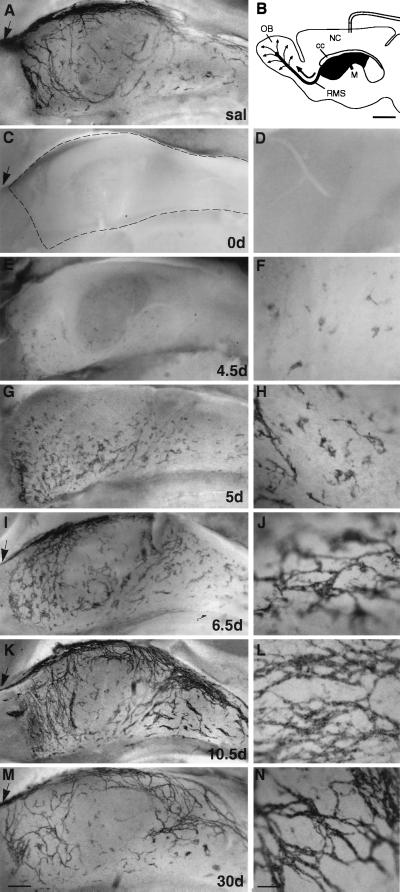
Figure 1.
Elimination and regeneration of the network of PSA-NCAM-positive chains in the SVZ. Mini-osmotic pumps containing Ara-C were implanted onto the surface of the right hemisphere as shown in B (scale bar: 1 mm). Low-magnification photomicrographs (A, C, E, G, I, K, and M; scale bar: 200 μm) are from the portion of the wall of the lateral ventricle indicated by dark gray in the schematic. (D, F, H, J, L, and N) Higher magnification (scale bar: 35 μm) for each of the time points. Dotted line in C outlines the borders of the lateral ventricle. Arrows indicate the RMS. (A) SVZ network of PSA-NCAM-positive chains in saline-infused mice. (C–N) Whole-mount PSA-NCAM immunostaining at different survivals after Ara-C treatment in the injected hemisphere. 0 days after Ara-C treatment, no PSA-NCAM immunostaining is found in SVZ (C and D). Individual PSA-NCAM-positive cells and small clusters first appear at 4.5 days scattered over the entire wall of the lateral ventricle (E and F). At 5 days the clusters are larger, and some short chains begin to form (G and H). By 6.5 days, many chains are now present and form a simple disorganized network (I and J). At 10 days a largely normal network is visible (K and L), although chains are more abundant than in saline-infused animals. At 30 days, the SVZ network appears normal (M and N). NC, neocortex; OB, olfactory bulb; cc, corpus callosum; M, Foramen of Monro.

