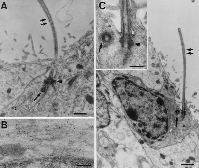
Figure 5.
(A) Some GFAP-positive type B cells contacting the ventricle exhibit a single cilium. After Ara-C treatment, type B cells contacting the ventricle extend a single cilium (double arrow). Characteristic basal bodies (arrowhead) and centriole (single arrow) are found at its base. (Scale bar: 0.25 μm.) (B) Postembedding immunostaining for GFAP revealed that this same cell has GFAP-positive intermediate filaments. The black dots overlying the thin intermediate filaments are gold particles. (Scale bar: 0.6 μm.) (C) In untreated mice, type B cells touching the ventricle with a single cilium (double arrow) also are found. (Scale bar: 1 μm.) (Inset) The characteristic basal bodies (arrowhead) and centriole (single arrow) associated with the base of the cilium. (Scale bar: 0.3 μm.)

