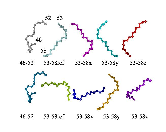
Figure 11.
Fragment alignment using RDC data for PF0255 from two different media. At the left of each line is the piece of the fragment before proline 53. The remaining four depictions of the piece after proline have been produced by rotating the reference structure by 180° about x, y, and z axes of the principal alignment frame. The structures in the second line have been rotated to overlay the first piece in both lines using the program chimera(Huang, 1996).

