
Figure 1.
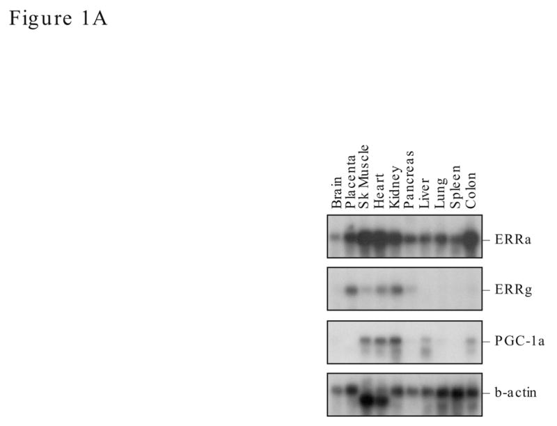
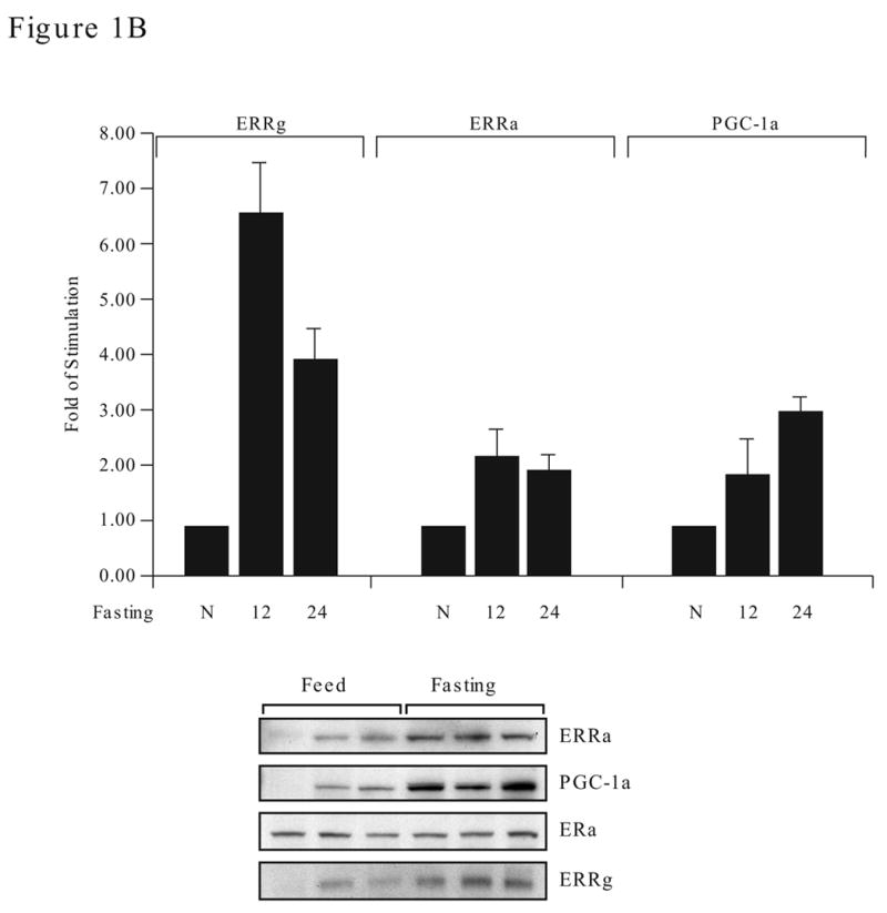
Expression of ERRα, ERRγ and PGC-1α in human tissues and mouse liver during fasting. A. Northern blot analyses of human tissues. ERRα, ERRγ, PGC-1α and β-actin probes (between 200–300 bp in length) were labeled to a similar specific activity and used in sequential hybridization to the FirstChoiceTM Northern Human Blot I from Ambion (2 μg polyA RNA /lane). The hybridization time for all four probes was 16 h, however, the exposure time varies. The x-ray film for ERRγ and PGC-1α were exposed for 48 h while ERRα was exposed for 24 h and β-actin for 2 h. B. ERRα, ERRγ and PGC-1α expression in mouse liver during fasting. Adult female mice (4 per group) were fed (N), fasted for 12 h (12) or 24 h (24). Total liver RNA was prepared from the individual mouse and analyzed by real time PCR with specific primers to ERRα, ERRγ or PGC-1α (upper). Data is presented as fold of stimulation from 4 mice ± SE. Liver nuclear protein lysates (30 μg protein) were individually prepared and the presence of ERRα, ERRγ, PGC-1α and ERα were detected by Western blot analyses with their respective antibodies (lower). The antibody dilution and exposure time as follow: ERRα, 1:1000 and 2 min; ERRγ, 1:200 and 1 min; PGC-1α, 1:500 and 5 min; ERα, 1:2000 and 5 min. There were three mice per experimental group and the length of fasting was 24 h.