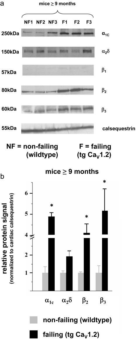
Figure 2. Protein expression of cardiac L-VDCC subunits in old wild-type and tg CaV1.2 mice.
(a) Specimens from old wild-type mice and tg CaV1.2 in heart failure were analyzed in immunoblots using specific polyclonal antibodies directed against the particular L-VDCC subunits.
(b) Protein expression of L-VDCC subunits was always normalized to cardiac calsequestrin protein expression in the same sample. Quantitative analysis of subunit protein expression is depicted as ratio of 10 months old tg CaV1.2 vs. age-matched wild-type. β1 protein bands were faint, and thus not analyzed quantitatively (number of WT/old tg CaV1.2 specimens was always identical for each subunit; n = 4). * p<0.05.

