
Figure 1.
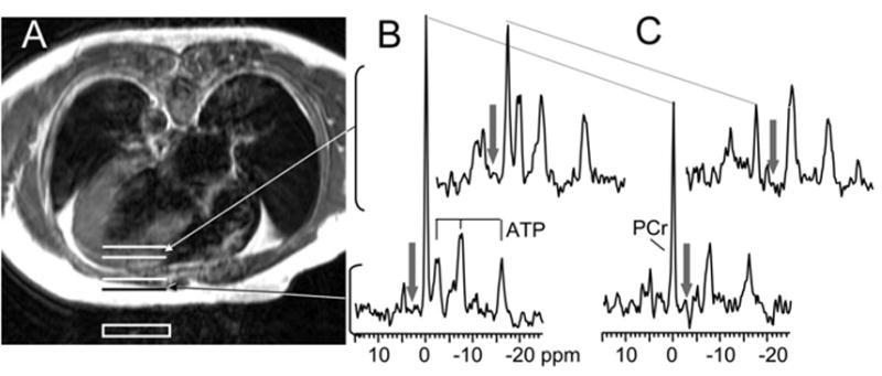
Axial spin-echo MR image (A) of a patient with LVH+CHF lying prone over a 31P surface coil (white box) and the corresponding localized 31P MR spectra from the chest (lower 2 spectra) and left ventricle (upper 2 spectra). The resonances derive from PCr and the γ-, α-, and β -phosphate resonances of ATP. The spectra were acquired with a 60° flip angle in the presence of chemically selective saturating irradiation (arrows) either in the control (B) or γ-ATP position (C). The decrease in the height of the PCr peak between control and γ-ATP saturation (dotted lines) is directly related to the rate of ATP synthesis through the CK reaction.