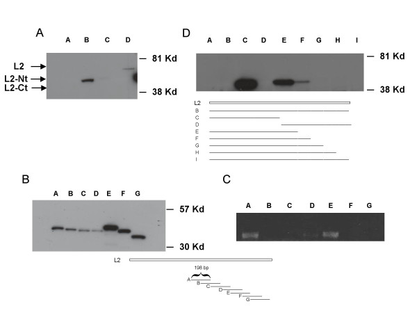
Figure 2.
Western analysis of HPV 31 L2. Cos-7 cells were transfected with GFP tagged L2 in pcDNA 3.1(-) expression vectors. After 72 hours, lysates were harvested and analyzed by Western blot with an antibody to GFP. (Panel A). Lane A: wild type HPV 31 L2. Lane B: wild type HPV 31 L2 N-terminus. Lane C: wild type HPV 31 L2 C-terminus. Lane D: codon optimized HPV 31 L2. (Panel B). Western analysis of transfections of GFP in frame fusions to 198 base pair fragments of the C-terminal domain of wild type L2. Each fragment overlapped with the adjacent fragment by 99 base pairs. Cell extracts were examined by Western analysis using a GFP antibody. Lane A: fragment A, GFP fusion to nucleotides 4771–4969. Lane B: fragment Bnucleotides 4870–5068. Lane C: fragment C, nucleotides4969–5167. Lane D: fragment D, nucleotides5068–5266. Lane E: fragment E, nucleotides5167–5365. Lane F: fragment F nucleotides 5266–5464. Lane G: fragment G, nucleotides5365–5568. (Panel C). RT-PCR analysis of RNAs isolated from cells transfected with plasmids shown in panel B. Primers to common GFP sequences were used in this analysis. (Panel D) Lane A: mock transfected cells. Lane B: complete wild type HPV 31 L2 fused to GFP nucleotides 4171–5568. Lane C: N-terminal domain wild type L2 (nucleotides 4171–4870) fusion to GFP. Lane D: C-terminal domain wild type L2 (nucleotides 4870–5568) fused to GFP. Lane E: N-terminal domain wild type L2 (nucleotides 4171–4969) fused to GFP. Lane F: N-terminal domain wild type L2 (nucleotides 4171–5068) fused to GFP. Lane G: N-terminal domain wild type L2 (nucleotides 4171–5166) fused to GFP. Lane H: N-terminal domain wild type L2 (nucleotides 4171–5265). Lane I: N-terminal domain wild type L2 (nucleotides 4171–5465). Cartoon identifies fragments of L2 fused to GFP examined in lanesE – I. Nucleotide numbers are those in HPV 31 genome.

