
Figure 2.
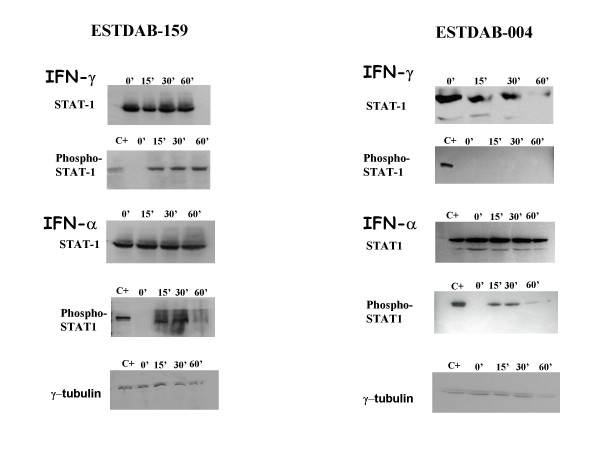
The expression and the activation of STAT proteins. Cells were treated with IFN-γ or IFN-α at 800 IU/ml. Proteins from cellular protein extracts were separated by SDS-PAGE and transferred to nitrocellulose membrane. Western blotting was performed as described in Materials and Methods. Anti-STAT1 and anti-phospho-STAT1 antibodies were used to assess STAT activation. Anti-γ-tubulin antibody was used to normalize the amounts of protein loaded in each well of the gel. Positive control (C+) of phosphorylated proteins from cellular extract A549 was obtained from Cell Signaling Technology.