Figure 10.

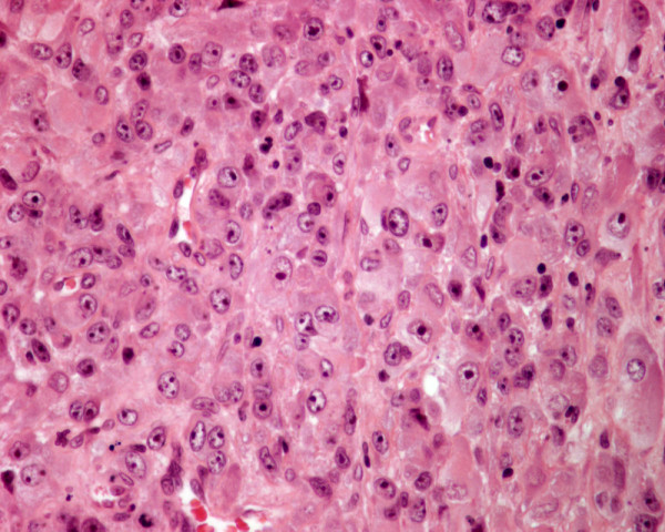
Histology of metastatic amelanotic melanoma to the thyroid in patient 4 shows large malignant polygonal cells with oval nuclei and prominent nucleoli, in solid pattern (hematoxylin and eosin, × 250).

Histology of metastatic amelanotic melanoma to the thyroid in patient 4 shows large malignant polygonal cells with oval nuclei and prominent nucleoli, in solid pattern (hematoxylin and eosin, × 250).