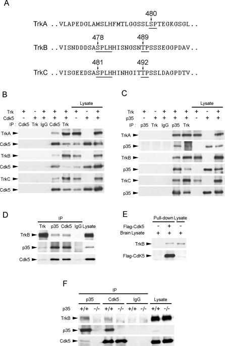
Figure 1. TrkB Interacted with Cdk5 and p35.
(A) TrkA, TrkB, and TrkC all contain proline-directed serine/threonine residues in the juxtamembrane region of the receptors (indicated by arrows). Nonetheless, only TrkB and TrkC contain Cdk5 consensus sites S/TPXK/H/R.
(B) Cell lysates from HEK293T cells overexpressing Cdk5 and TrkA, TrkB, or TrkC were immunoprecipitated (IP) with Cdk5 antibody and immunoblotted with pan-Trk antibody. TrkA, TrkB, and TrkC were all observed to associate with Cdk5.
(C) Cell lysates from HEK293T cells overexpressing p35 and TrkA, TrkB, or TrkC were immunoprecipitated with p35 antibody and immunoblotted with pan-Trk antibody. TrkA, TrkB, and TrkC were all observed to associate with p35.
(D) Brain lysate from P7 rat brain was immunoprecipitated with pan-Trk, p35, or Cdk5 antibody and immunoblotted with p35, Cdk5, and TrkB antibodies. Rabbit normal IgG was used as a control. TrkB was observed to associate with both p35 and Cdk5 in P7 rat brain.
(E) The membrane fraction of adult brain lysates was incubated with or without Flag-tagged Cdk5. Flag-tagged Cdk5 pulled down TrkB from the membrane fraction of adult brain lysates.
(F) Brain lysates from P7 p35+/+ or p35−/− mouse brains were immunoprecipitated with p35 and Cdk5 antibodies and immunoblotted with p35, Cdk5, and TrkB antibodies. Rabbit normal IgG served as a control. Association between Cdk5 and TrkB was abolished in p35−/− brain, indicating that p35 was required for the association between Cdk5 and TrkB.

