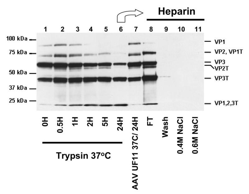
Fig. 4. Mildly trypsinized but not fully trypsinized virions bind heparin.

AAV2-GFP purified by trypsin/DOC/CsCl/heparin (lane 1) was further treated with 0.02% trypsin for 30 minutes (lane 2), 1 hour (lane 3), 2 hours (lane 4), 5 hours (lane 5), or 24 hours (lane 6). As a control, the purified virus from lane 1 was incubated for 24 hours at 37°C without trypsin (lane 7). Virus treated for 24 hours with trypsin (lane 6) was loaded onto a heparin affinity column: Lane 8, flow-through; Lane 9, wash; Lane 10, elution with 0.4M NaCl; Lane 11, elution with 0.6M NaCl. Capsid proteins were probed with anti-AAV2 polyclonal antisera. The position of the tryptic fragments are given on the right as indicated in Figure 2.
