
Fig. 5. Proteolysis distinguishes full and empty AAV2 particles. A. Trypsin.
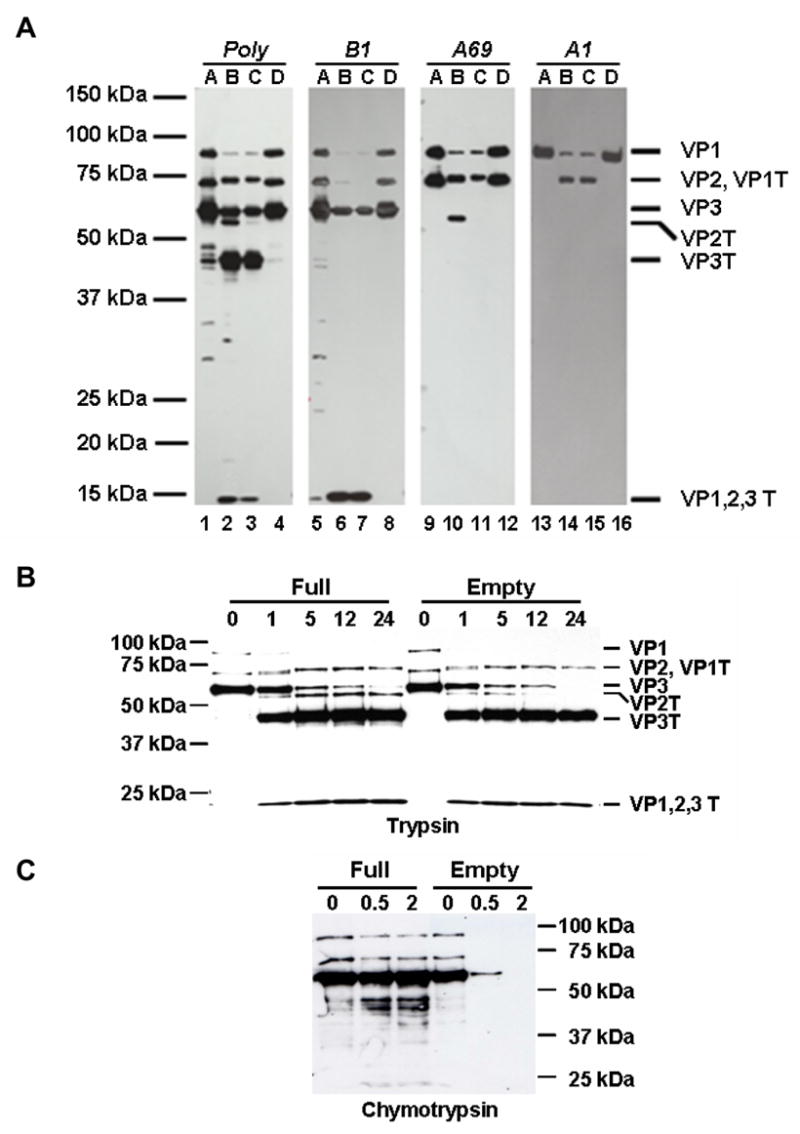
Purified full (F-T/iodixanol/heparin) and empty AAV2 capsids were treated with 0.02% trypsin, and the capsid proteins were probed with the indicated antibodies (AAV2 polyclonal, A1, B1, A69). Lanes A, AAV2 full capsids; lanes B, AAV2 full capsids digested with trypsin for 24 hours; lanes C, AAV2 empty capsids digested with trypsin for 24 hours; lanes D, AAV2 empty capsids. B. Trypsin time course. Purified full (F-T/iodixanol/heparin) and empty AAV2 virions were treated with trypsin for the time (hours) indicated above each lane and probed with anti-AAV2 polyclonal antisera. C. Chymotrypsin time course. Purified full (F-T/iodixanol/heparin) and empty AAV2 capsids were treated with chymotrypsin for the time (hours) indicated above each lane and the capsid proteins were probed with anti-AAV2 polyclonal antisera.