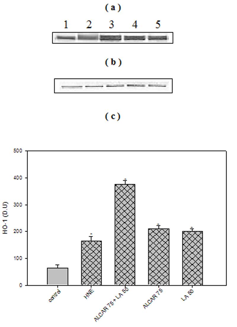
Fig. 4.

ALCAR+LA-mediated up-regulation of HO-1. (a) Representative Western immunoblot analysis of neuronal cells for HO-1 protein. 100μg of protein were analysed by sodium dodecyl sulphate–polyacrylamide gel electrophoresis and immunoblotting using a mouse monoclonal anti-HO-1 antibody. The grid bars are in presence of HNE and the plain bars are without HNE. Lane 1: control (untreated cells); Lane 2: 10μM HNE; Lane 3: 10 μM HNE+75μM ALCAR+50μM LA; Lane 4: 75μM ALCAR+10μM LA; Lane 5: 10μM HNE+50 μΜ LA (b) Anti-GAPDH blot as control for equal protein loading. (c) Densitometric analysis from five independent experiments (mean ±SD of values expressed as relative units). Significant differences were assessed by ANOVA. Post hoc analysis was via Student-Newman-Keuls test, and the P values given are compared with the control. *p < 0.005.
