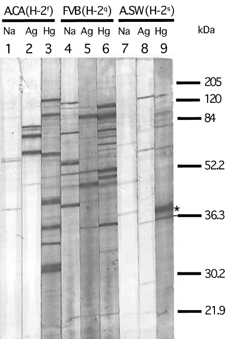
Fig 4.
Immunoblotting of sera from mercury- and silver-treated A.CA (H-2f), FVB/N (H-2q) and A.SW (H-2s) mice using nuclear extract from Hep-2 cells. Nitrocellulose strips blotted with a Hep-2 nuclear extract were incubated with a representative serum (at 1 : 100 dilution) from an A.CA (H-2f), a FVB/N (H-2q) and an A.SW (H-2s) mouse injected with saline (lanes 1, 4 and 7), silver (lanes 2, 5 and 8) and mercury (lanes 3, 6 and 9) as described in Fig. 3. Thereafter, the bound IgG antibodies were detected with alkaline phosphatase-conjugated goat antimouse IgG1 (see for further description). A relatively strong staining of a 36-kDa band corresponding to fibrillarin (as indicated by an underlined asterisk) can be observed in all mercury-treated mice (lanes 3, 6 and 9). Sera from silver-treated FVB/N (H-2q) and A.SW (H-2s) mice also show a weak staining of the same band (lanes 5 and 8). However, the serum from the silver-treated mouse A.CA (H-2f) shows no staining of the corresponding band (lane 2).

