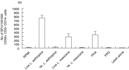
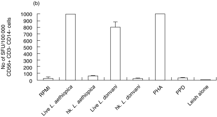
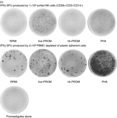
Fig. 2.
Leishmania induced IFNγ secretion in NK cells purified from human PBMC by FACS (two donors tested D18 and D6). Graphs show IFNγ spot forming units (SFU) in cells (a) D18 cells sorted for CD56+/CD3-, CD14- after 40 h stimulation and (b) D6 cells sorted for CD56+/CD3– after 48 h stimulation. Bars show mean SFU of three wells counted + SD. Spot frequencies greater than ≥1000 SFU/well are represented as 1000 SFU. (c)Representative picture of Leishmania aethiopica induced IFNγ SFU produced by FACS purified NK cells (D18), pictures of SFU produced by PBMC are shown as comparison.


