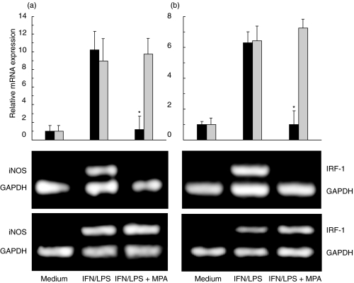
Fig. 4.
MPA inhibits iNOS and IRF-1 gene expression in fibroblasts. Primary fibroblasts (black bars, and upper photographs) and macrophages (grey bars, and lower photographs) were cultured in medium alone, or stimulated with IFN-γ (250 U/ml) + LPS (5 µg/ml), in the presence or absence of MPA (5 µm). The photographs of the gels from a representative of three separate experiments are presented. The results of RT-PCR analysis, presented as fold increase in iNOS (a) or IRF-1 (b) gene expression relative to the expression in untreated cells, are means ± s.d. of three independent experiments (*P < 0·01 refers to IFN/LPS treatment).

