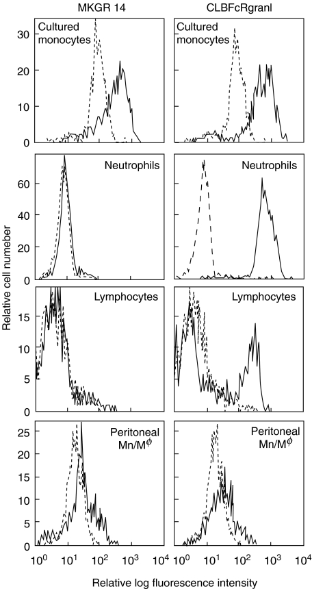
Fig. 1.
Histograms of anti-FcγRIII MoAb MKGR14 binding cells. Four-day-cultured monocytes, peripheral blood leucocytes (NA (1 +, 2 +) donor) or peritoneal cells were incubated on ice with anti-FcγRIII MoAb MKGR14 (left) or CLBFcRgranI (right). Neutrophils, lymphocytes or peritoneal monocytes/macrophages were identified by their characteristic forward and side scatter properties, and the cell population exhibiting these characteristics was selected by flow cytometric gating for analysis. The black lines indicate anti-FcγRIII MoAb binding cells and the grey lines indicate negative control.

