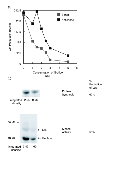
Fig. 5.
Blocking Lck protein synthesis and kinase activity results in accelerated viral replication. (a) Jurkat T cells (106/ml) were untreated or incubated with 1–5 µm phosphorothioate oligodeoxynucleotide 15mer antisense-lck or sense-lck sequences 12–16 h prior to infection with HIV-1IIIB (moi 1·5). Viral gag p24 antigen production was then measured by ELISA after 2 days of culture. Results represent the average of duplicate cultures. These results are representative of at least three independent experiments. (b) The level of newly synthesized Lck protein and Lck kinase activity following treatment with antisense-lck or sense-lck 15mer phosphorothioate oligodeoxynucleotide sequences as in A, above. Newly synthesized Lck was assessed by specific immunoprecipitation using anti-Lck after metabolic labelling with [35S]-labelled amino acids. The [35S]-labelled Lck was isolated by SDS-PAGE and the protein visualized by autofluorography. The kinase activity of total cellular Lck was assessed by in vitro kinase assay using rabbit muscle enolase as an exogenous substrate (see). The percentage reduction in newly synthesized Lck protein and total cellular Lck kinase activity of the antisense compared to the sense treated cells was determined after densitometry analysis of the autofluorograph and autoradiograph, respectively.

