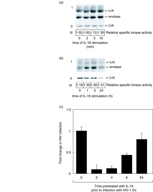
Fig. 7.
IL-16 stimulation results in a sustained increase in Lck kinase activity that correlates with inhibition of HIV-1 infection. (a) In vitro kinase assay (I) and Western immunoblot (II) of Lck protein following stimulation of Jurkat T cells (106/ml) with 5 × 10⁻⁹m human recombinant IL-16 for 2–10 min Arrows indicate location of Lck and enolase. The relative specific kinase activity of Lck (III) was determined as previously described [14] as the ratio of the optical density of the enolase band measured from the kinase experiment to the optical density of the Lck protein band measured from the Western immunoblot. (b) In vitro kinase assay (I) and Western immunoblot (II) of Lck protein following stimulation of Jurkat T cells (106/ml) with 5 × 10⁻⁹M human recombinant IL-16 for 1–24 h. Arrows indicate the position of Lck and enolase. The relative specific kinase activity (III) of Lck was determined as described above. These results are representative of at least three independent experiments. C. HIV-1 viral replication as measured by gag p24 ELISA following IL-16 treatment of Jurkat T cells. Jurkat cells (106/ml) were untreated or pretreated with 5 × 10⁻⁹M human recombinant IL-16 for the indicated times prior to infection with HIV-1IIIB (moi 0·3). Results represent the mean p24 antigen level ± SEM of triplicate cultures. These results are representative of at least three independent experiments.

