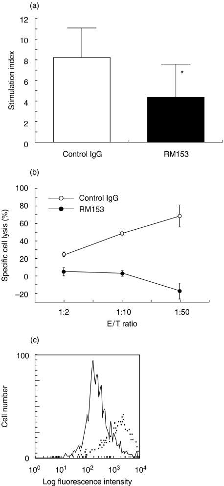
Fig. 4.
Co-stimulatory activity of CD30/CD30L interaction in T cell proliferation and β cell destruction. (a) Splenic T cells were cultured with MMC-treated NOD islets in the presence of control IgG (□) or RM153 (▪). After 3 days, 1 µCi 3H-thymidine was added 16 h before harvesting. Data are presented as mean ± SD stimulation index of triplicated samples. *P < 0·05 versus control. (b) Specific lysis by islet-derived CD8+ CTL in the presence of anti-CD30L mAb (•) or control IgG (○) was assessed by 8 h 51Cr-release assay at the indicated E/T ratios. One representative of three separate experiments was shown (mean ± SE). (c) Expression of CD30L on NOD islet cells was evaluated after exposure to IFN-γ. Freshly isolated islet cells from NOD mice were cultured with 100 ng/ml IFN-γ for 24 h and then were stained with biotin-conjugated anti-CD30L mAb (………) and FITC-conjugated-avidin (——).

