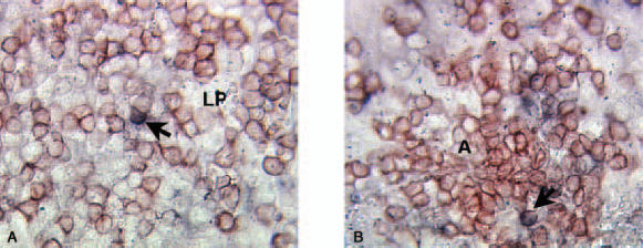
Fig. 5.

Immunohistochemical double staining of IL-10 (blue) and CD3 (red) in the colonic mucosa of UC patient. (a) A double positive cell (thick arrow) in the lamina propria outside basal lymphoid aggregate among several single stained CD3+ cells. (b) A double positive cell (thick arrow) in the T cell area of a basal lymphoid aggregate. For abbreviations see Fig. 4. Original magnification ×220.
