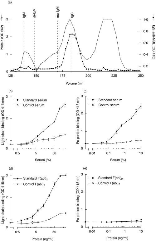
Fig. 3.
Biochemical characterization of anti-mannose-binding lectin (anti-MBL) autoantibodies. After gel filtration, on Superdex HR 200, of serum from a patient with systemic lupus erythematosus (SLE), fractions were analysed for the presence of immunoglobulin (Ig)G anti-MBL (indicated by the black circles). The protein pattern, as well as the position of filtration of IgM, dimeric IgA (di-IgA), monomeric IgA (mo-IgA) and IgG are depicted (a). Anti-MBL reactivity was detected in serial dilutions of whole serum (b) and (c), and purified F(ab′)2 fragments (d) and (e), using antibodies against the light chains (b) and (d) and the Fc portion (monoclonal antibody HB43) (c) and (e), respectively.

