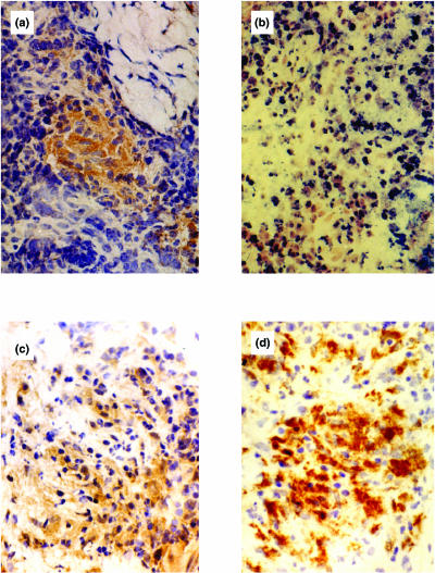
Fig. 1.
Immunohistochemical detection of chemokine and receptor protein in borderline tuberculoid leprosy in reaction (BT-RR) leprosy skin lesions. (a) MCP-1 chemokine-protein-positive cells within a granuloma. (b) MCP-1 chemokine-mRNA positive staining by in situ hybridization. (c) A section of the same lesion stained with the polyclonal antibody CCR2 (a chemokine receptor for MCP-1). (d) Immunostaining of the same skin lesion with the monoclonal antibody CD68 (macrophage marker) indicating a large proportion of positive cells.

