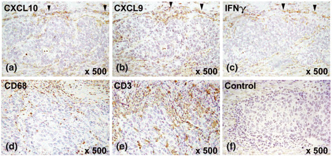
Fig. 4.
CXCL10, CXCL9 and IFN-γ expression in gastric carcinoma by immunohistochemistry. The figure shows examplarily an intestinal-type gastric carcinoma. CXCL10 (a) and CXCL9 (b) were expressed by mononuclear cells, which infiltrate gastric carcinoma in areas with IFN-γ production (c). Expression was most pronounced in close proximity to the expanding tumour border (marked by arrowheads). Tumour cells themselves did not express CXCL10 and CXCL9. Serial sections stained with CD3 and CD68 identified cells at the tumour border as macrophages (d) and T cells (e). The negative control showed no staining of the mononuclear cells at the tumour border. Magnification (a–f) ×500.

